Abstract
Arboviral infections represent an expanding global public health threat, with increasing recognition of their neurological complications. Dengue, chikungunya, Zika, yellow fever, and West Nile viruses are now well-established causes of central and peripheral nervous system disease. Neurological manifestations range from transient encephalopathy and cognitive impairment to encephalitis, myelitis, acute flaccid paralysis, movement disorders, immune-mediated neuropathies, and congenital neurodevelopmental abnormalities. Advances in epidemiology, neuroimaging, and experimental virology have challenged the traditional view that several of these viruses are non-neurotropic. This review synthesizes contemporary epidemiologic, clinical, and mechanistic data on arboviral neurological disease, with particular emphasis on flaviviruses, and highlights implications for diagnosis, clinical management, and future research.
Highlights
- Many more arboviruses are increasingly being recognized as neurotropic. Viruses including dengue and chikungunya—once considered non-neurotropic—are now well-documented causes of serious central and peripheral nervous system disease.
- Blood–brain barrier disruption is a central mechanism of injury. These viruses can cross into the CNS either as free viral particles or via infected immune cells (a Trojan horse mechanism), triggering inflammation that breaks down tight junctions in the BBB and allows for further neurological damage.
- Each arbovirus carries a distinct neurological profile. Dengue causes encephalopathy, movement disorders, and stroke; Zika is strongly linked to Guillain–Barré syndrome through molecular mimicry, as well as congenital neurodevelopmental abnormalities; chikungunya produces acute encephalitis and myelitis alongside chronic arthralgias and inflammatory arthritis that can persist for months to years; West Nile virus is a leading cause of neuroinvasive arboviral disease.
- Climate change and global travel are expanding arboviral risk worldwide. Geographic overlap in environmental suitability for dengue, chikungunya, Zika, and yellow fever is increasing, raising the likelihood of co-circulation in new regions and compounding public health impact—making awareness of neurological complications critical for clinicians in both endemic and emerging areas.
Introduction
Arboviruses, or arthropod-borne viruses, are transmitted primarily through mosquito and tick vectors and are responsible for substantial global morbidity and mortality. Dengue, chikungunya, Zika, yellow fever, and West Nile viruses account for most of clinically significant human diseases and continue to expand geographically as a result of climate change, urbanization, and global travel. Recent modeling studies demonstrate extensive overlap in the global environmental suitability of dengue, chikungunya, Zika, and yellow fever viruses, increasing the likelihood of co-circulation and compounded public health impact [1]. Although many arboviral infections are self-limited, neurological involvement is increasingly recognized and contributes disproportionately to severe disease, long-term disability, and mortality [2–6].
Historically, several arboviruses, particularly dengue virus, were considered non-neurotropic, and neurological manifestations were attributed to metabolic derangements or systemic illness. However, accumulating clinical, epidemiologic, and experimental evidence has demonstrated that neurological complications are both real and clinically significant. Large population-based studies, updated reviews, and mechanistic investigations now describe a wide spectrum of central and peripheral nervous system manifestations associated with arboviral infections, necessitating heightened awareness among neurologists and infectious diseases clinicians practicing in endemic and emerging regions [2–7].
The impact of arboviral infection on the nervous system may involve both the central and peripheral nervous systems. Some viruses demonstrate more profound effects on the PNS compared to the CNS, but classification of arboviral neurologic effects based on site can be difficult to quantify. Zika virus is more classically associated with Guillain–Barré syndrome compared to West Nile virus, while dengue and, to a lesser extent, chikungunya are known to cause PNS disease with GBS [8].
Mechanisms of Neurological Injury
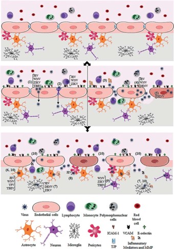
Neurological complications of arboviral infections arise through a complex interplay of direct viral neuroinvasion, disruption of the blood–brain barrier (BBB), immune-mediated injury, and vascular dysfunction (Figure 1). Central to many of these processes is impairment of the BBB, a highly specialized interface that regulates central nervous system homeostasis [2].
The intact BBB is composed of endothelial cells tightly adhered through tight junction and adherens junction proteins and functioning in close association with pericytes, astrocytic end-feet, and microglia. Together, these elements form the neurovascular unit, which tightly controls the flux of solutes, circulating immune cells, and pathogens from the bloodstream into the central nervous system [2].
Flaviviruses may cross the BBB as free viral particles or via infected leukocytes through a Trojan horse mechanism. Systemic inflammation and direct infection of brain microvascular endothelial cells induce downregulation of tight junction proteins, endothelial dysfunction, and increased permeability, facilitating viral entry into the CNS [2].
Once within the CNS, arboviruses may infect astrocytes, microglia, pericytes, and neurons. Activation of astrocytes and microglia induces the release of inflammatory mediators, cytokines, chemokines, and matrix metalloproteinases, further disrupting BBB integrity and amplifying neuronal injury [2].
Molecular mimicry leading to post-infectious autoimmune neuropathy, primarily GBS, arises through immune cross-reactivity between arboviral antigens and peripheral nerve epitopes. In ZIKV infection, axonal transport of virus from cutaneous small fibers to peripheral nerves may additionally provide a direct pathway for peripheral nerve injury independent of the immune-mediated mechanism. Acute disseminated encephalomyelitis (ADEM) represents the CNS counterpart of this immune-mediated process, driven by molecular mimicry affecting central myelin. Both GBS and ADEM may occur following infection with ZIKV, CHIKV, or DENV [8,9].
Patterns of Neuroinvasive Effects Based on Arbovirus Infection
The below section highlights specific neuroinvasive diseases based on infections with dengue, chikungunya, Zika and West Nile viruses [Table 1].
Dengue Virus
Dengue virus demonstrates neurotropic potential through direct neuroinvasion, systemic inflammatory effects, vascular injury, and post-infectious immune-mediated mechanisms. Encephalopathy occurs in approximately 5% of hospitalized cases and reflects systemic complications including hepatic dysfunction, metabolic derangements, hypotension, and cytokine-mediated endothelial injury. Severe cases may be complicated by cerebral edema, intracranial hemorrhage, and ischemic stroke [3,4].
Dengue encephalitis represents direct CNS infection and accounts for approximately 27% to 39% of dengue-associated neurological cases. Detection of viral antigens and RNA in brain tissue and cerebrospinal fluid supports direct neuroinvasion. Dengue virus infects brain microvascular endothelial cells, inducing BBB disruption [2–4].
Movement disorders including tremor, ataxia, and dystonia typically develop within 14 days of infection and correspond to basal ganglia or brainstem abnormalities. Memory loss and cognitive impairment have been described, with hippocampal involvement demonstrated on imaging [3,4].
Population-based data demonstrate increased odds of new-onset neurological disease, memory loss, and movement disorders within 30 to 90 days after dengue infection, particularly among individuals older than 69 years and during periods of dengue virus serotype 3 transmission. Absolute event rates remain low [7]. Immune-mediated syndromes including acute disseminated encephalomyelitis, cerebellitis, Guillain–Barré syndrome, and isolated cranial neuropathies have been reported [3,4].
Chikungunya Virus
Chikungunya virus causes neurological disease through direct viral neuroinvasion, cytopathic effects, and immune-mediated injury. Viral RNA has been detected in cerebrospinal fluid, confirming direct CNS infection. Activation of astrocytes and microglia amplifies inflammatory mediator release and BBB disruption [5].
Neurological manifestations of chikungunya virus infection include encephalitis with confusion, hyperreflexia, and sensory level abnormalities; prominent myelitis; acute disseminated encephalomyelitis; Guillain–Barré syndrome and atypical variants; seizures; myoclonus; altered mental status; and post-infectious cognitive dysfunction [5]. In addition to these neurologic complications, chikungunya is uniquely characterized by a high burden of persistent musculoskeletal symptoms, with many patients developing chronic arthralgias, myalgias, and inflammatory arthritis that may persist for months to years after acute infection. These chronic manifestations are thought to reflect ongoing immune-mediated inflammation and viral persistence within joint and periarticular tissues, and they frequently coexist with or exacerbate neurologic disability, contributing substantially to long-term functional impairment and reduced quality of life.
Zika Virus
Zika virus affects both the peripheral and central nervous systems through distinct but overlapping pathogenic mechanisms. Peripheral nervous system involvement is predominantly immune-mediated, with neurological symptoms typically developing 5 to 10 days after the acute viral illness. Guillain–Barré syndrome is the most frequently reported neurologic complication and encompasses several subtypes, including acute inflammatory demyelinating polyneuropathy, acute motor axonal neuropathy, and Miller–Fisher syndrome. These manifestations are believed to arise from molecular mimicry, with the generation of antiganglioside autoantibodies that cross-react with peripheral nerve components, leading to immune-mediated nerve injury. In addition to classic Guillain–Barré syndrome, patients may develop cranial neuropathies, autonomic dysfunction, and prolonged weakness, underscoring the potential for significant morbidity even after resolution of the initial infection [2,6].
Zika virus exhibits marked tropism for neural progenitor stem cells during fetal development, a property that underlies its devastating effects on the developing central nervous system. Direct infection of these cells interferes with cell-cycle progression, induces apoptosis, and impairs neuronal differentiation, thereby disrupting normal neurogenesis and cortical development. These pathogenic processes result in microcephaly and the constellation of abnormalities collectively termed congenital Zika syndrome, which includes cortical thinning and malformations, ventriculomegaly, subcortical and intracranial calcifications, ocular defects such as chorioretinal atrophy and optic nerve hypoplasia, and profound, often irreversible neurodevelopmental impairment. Affected infants frequently demonstrate severe cognitive delay, seizures, spasticity, feeding difficulties, and long-term motor dysfunction, highlighting the enduring neurologic burden of in utero Zika virus infection. In addition to direct neuronal injury, infection of brain microvascular endothelial cells and activation of astrocytes and microglia further compromise blood–brain barrier integrity, amplifying neuroinflammation and contributing to the severity of congenital disease [2,6].
West Nile Virus
West Nile virus is a leading cause of arboviral neuroinvasive disease. Neuroinvasion occurs via hematogenous spread, BBB disruption, infection of brain microvascular endothelial cells, and leukocyte trafficking [2,6].
Clinical manifestations of West Nile virus infection span a broad neurologic spectrum and include meningitis, encephalitis, acute flaccid myelitis, and a variety of movement disorders. Acute flaccid myelitis is a distinctive and severe manifestation resulting from direct viral injury to anterior horn cells of the spinal cord, leading to rapid onset of asymmetric flaccid weakness, hyporeflexia or areflexia, and, in severe cases, involvement of respiratory musculature necessitating ventilatory support. Recovery is frequently incomplete, with many patients experiencing persistent motor deficits [2,6].
Encephalitis represents the most common form of neuroinvasive disease and may be complicated by seizures, altered mental status, and focal neurologic deficits. Movement disorders, including tremor, parkinsonism, and cerebellar ataxia, reflect viral tropism for the basal ganglia, thalamus, and brainstem. Additional manifestations include cranial neuropathies, brachial plexopathy, and ocular involvement such as chorioretinitis and uveitis. Post-infectious immune-mediated complications, including Guillain–Barré syndrome, have also been reported and contribute to prolonged morbidity, particularly among older adults and individuals with underlying comorbidities [6].
Conclusions
Arboviral infections are increasingly recognized as important and often underappreciated causes of neurologic disease worldwide. Dengue, chikungunya, Zika, yellow fever, and West Nile viruses can produce a wide spectrum of central and peripheral nervous system manifestations through overlapping mechanisms that include direct neuroinvasion, blood–brain barrier disruption, immune-mediated injury, and vascular dysfunction [Table 1]. Increasing epidemiologic and experimental data have firmly established that several arboviruses previously considered non-neurotropic, particularly dengue virus, can cause clinically meaningful neurologic complications [Table 2].
The clinical phenotypes associated with arboviral infection are heterogeneous and influenced by viral tropism, host immune response, age, and comorbid conditions. Older adults appear to be at increased risk for severe neurologic outcomes, including encephalitis, movement disorders, and cognitive impairment, while immune-mediated complications such as Guillain–Barré syndrome may occur across age groups. Recognition of these patterns is critical, as neurologic manifestations may develop during acute infection or emerge in the subacute post-infectious period, often in the absence of classic systemic features.
Timely diagnosis of arboviral neurologic disease remains challenging due to overlapping clinical syndromes, limited early diagnostic sensitivity, and variable access to confirmatory testing. As geographic expansion and co-circulation of arboviruses continue, heightened clinical awareness and careful epidemiologic assessment will be essential for early recognition and appropriate management. Future research priorities include improved diagnostic strategies, a deeper understanding of viral and host determinants of neurotropism, and development of targeted preventive and therapeutic interventions to mitigate the growing neurologic burden of arboviral infections.
| Virus | Mechanisms of Injury | Major Neurological Syndromes |
|---|---|---|
| Dengue virus | BBB disruption; direct CNS invasion; systemic inflammatory and vascular injury; immune-mediated mechanisms | Encephalopathy; encephalitis; movement disorders; cognitive impairment; ADEM; Guillain–Barré syndrome |
| Chikungunya virus | Direct viral neuroinvasion; glial activation; immune-mediated injury | Encephalitis; myelitis; ADEM; Guillain–Barré syndrome; seizures |
| Zika virus | Immune-mediated peripheral neuropathy; neural progenitor cell infection; BBB disruption | Guillain–Barré syndrome; congenital Zika syndrome; encephalitis |
| West Nile virus | BBB disruption; direct neuronal infection; immune-mediated injury | Encephalitis; meningitis; acute flaccid myelitis; movement disorders |
| Neurological Syndrome | Commonly Associated Viruses | Key Clinical Features |
|---|---|---|
| Encephalopathy | Dengue | Occurs during acute systemic illness; metabolic and vascular contributors |
| Encephalitis | Dengue, Chikungunya, West Nile, Zika | Altered mental status, seizures, focal deficits |
| Acute flaccid myelitis | West Nile | Asymmetric weakness, areflexia, potential respiratory failure |
| Myelitis | Chikungunya, Dengue | Acute motor and sensory deficits |
| Movement disorders | Dengue, West Nile | Tremor, parkinsonism, cerebellar ataxia |
| Guillain–Barré syndrome | Zika, Dengue, Chikungunya | Post-infectious ascending weakness |
| Congenital neurodevelopmental disease | Zika | Microcephaly, cortical malformations |
Funding
This research received no external funding.
Acknowledgments
The author used Claude for content assistance, specifically to create an initial draft from prior personal presented material. The manuscript was reviewed and revised and the author takes full responsibility for the content of this publication.
Conflicts of Interest
The author declares no conflict of interest.
References
- Lim, A.; Shearer, F.M.; Sewalk, K.; Pigott, D.M.; Clarke, J.; Ghouse, A.; Judge, C.; Kang, H.; Messina, J.P.; Kraemer, M.U.; et al. The overlapping global distribution of dengue, chikungunya, Zika and yellow fever. Nat. Commun. 2025, 16, 3418. [CrossRef] [PubMed]
- Mustafá, Y.M.; Meuren, L.M.; Coelho, S.V.A.; de Arruda, L.B. Pathways Exploited by Flaviviruses to Counteract the Blood-Brain Barrier and Invade the Central Nervous System. Front. Microbiol. 2019, 10, 525. [CrossRef] [PubMed]
- Carod-Artal, F.J.; Wichmann, O.; Farrar, J.; Gascón, J. Neurological complications of dengue virus infection. Lancet Neurol. 2013, 12, 906–919. [CrossRef] [PubMed]
- Fong, S.L.; Wong, K.T.; Tan, C.T. Dengue virus infection and neurological manifestations: An update. Brain 2024, 147, 830–838. [CrossRef] [PubMed]
- Supanchana, P.; Kirushanth, C.; Wijewardana, C.T.; Peiris, L.T.; Jayasinghe, J.; Rudra, A.; Govindapala, D.; Higgoda, R.; Thivakaran, T.; Muthugala, R.; et al. A case series of neuro-chikungunya: Unmasking the neurotropic potential of chikungunya virus. BMC Infect. Dis. 2025, 26, 32. [CrossRef] [PubMed]
- Gould, C.V.; Staples, J.E.; Guagliardo, S.A.J.; Martin, S.W.; Lyons, S.; Hills, S.L.; Nett, R.J.; Petersen, L.R. West Nile Virus: A Review. JAMA 2025, 334, 618–628. [CrossRef] [PubMed]
- Wee, L.E.; Tan, W.Z.; Chow, J.Y.; Lim, J.T.; Chiew, C.; Chia, P.Y.; Dickens, B.; Ng, L.C.; Ong, B.; Leo, Y.S.; et al. Neurological Events Associated with Acute Dengue Infection. JAMA Neurol. 2026, 83, 171–180. [CrossRef] [PubMed]
- Cao-Lormeau, V.M.; Blake, A.; Mons, S.; Lastère, S.; Roche, C.; Vanhomwegen, J.; Dub, T.; Baudouin, L.; Teissier, A.; Larre, P.; et al. Guillain-Barré Syndrome outbreak associated with Zika virus infection in French Polynesia: A case-control study. Lancet 2016, 387, 1531–1539. [CrossRef] [PubMed]
- Corrêa, D.G.; Freddi, T.A.L.; Chaves, C.G.; Hygino da Cruz, L.C., Jr. Neuroimaging features of arboviral infections in the Americas. Clin. Imaging 2022, 85, 64–73. [CrossRef] [PubMed]