
Abstract
Central nervous system (CNS) blastomycosis manifesting as hypophysitis is an exceptionally rare fungal presentation that can mimic other sellar masses, posing significant diagnostic challenges in immunocompetent individuals. A woman in her early 20s with atopic dermatitis previously on dupilumab presented with headaches, diplopia, and blurred vision, with magnetic resonance imaging (MRI) revealing a sellar/suprasellar mass. Initial surgical pathology demonstrated granulomatous inflammation, which led to empirical treatment with corticosteroids and infliximab for presumed neurosarcoidosis. Following progressive neurologic decline, central diabetes insipidus, and cerebral infarcts, cerebrospinal fluid (CSF) cultures grew Blastomyces dermatitidis six weeks into her hospitalization. The patient was treated with one month of liposomal amphotericin B followed by a year and three months of voriconazole, resulting in substantial resolution of her neurologic and endocrine symptoms, normalization of CSF pleocytosis, and clearance of urine antigens. Ultimately, blastomycosis-associated hypophysitis must be considered in the differential diagnosis of sellar lesions, particularly in patients from endemic regions like Illinois. Furthermore, clinicians must recognize that empirical corticosteroid and infliximab therapy may contribute to rapid progression of an unrecognized fungal infection. This case underscores the necessity of maintaining a broad differential and pursuing prolonged diagnostic evaluation to avoid dangerous misdiagnoses, enable timely targeted antifungal therapy, and prevent permanent complications.
1. Introduction
Blastomycosis is an uncommon systemic fungal infection caused by the thermally dimorphic fungi Blastomyces dermatitidis and Blastomyces gilchristii, which are endemic to the Ohio and Mississippi River valleys and the Great Lakes region [1]. Reported incidence is generally low but varies by geography. Minnesota and Wisconsin report higher statewide rates, with substantially higher incidence in some hyperendemic areas [1,2]. Reported cases in Illinois increased from 24 in 1993 to 87 in 2002 [3]. However, more recent epidemiologic data are limited, as blastomycosis is not currently a reportable disease in Illinois [4]. This lack of mandatory reporting likely limits current population-level surveillance and incidence estimates. This may obscure the true burden of disease in the region, especially given that blastomycosis can clinically mimic other granulomatous processes such as tuberculosis or sarcoidosis [5,6].
Blastomyces spp. exist in the environment as molds that produce infectious spores which become airborne following disruption of contaminated soil. Inhalation of these spores is the primary route of infection, with the lungs being the most involved site in 79% of cases [7]. CNS involvement is rare, occurring in 5–10% of disseminated infections [7], and pituitary involvement is even more uncommon, with only isolated cases being reported [8–11]. CNS spread is believed to occur via lymphohematogenous dissemination [12,13].
We report a diagnostically challenging and rare case of CNS blastomycosis manifesting as hypophysitis in a previously healthy young woman, initially presumed to have neurosarcoidosis.
2. Case Description
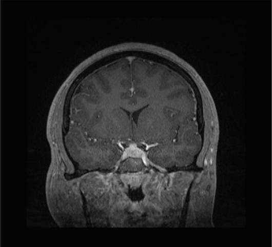
A woman in her early twenties with a medical history of atopic dermatitis (previously on dupilumab) presented to the emergency department with three months of headaches and three weeks of diplopia and blurred vision. MRI showed a 2 × 2 cm sellar/suprasellar mass concerning for a pituitary macroadenoma, with slight optic chiasm compression, suprasellar extension, and involvement of the right cavernous sinus (Figure 1). Chest imaging also demonstrated right-sided pneumonia. A subsequent bronchoscopy was performed, which yielded a negative microbiological workup. While hospitalized, she developed bilateral cranial nerve VI palsies and underwent transsphenoidal exploration for the presumed pituitary macroadenoma. Intraoperatively, no discrete adenoma was identified; the gland appeared diffusely inflamed and beefy-red, raising concern for a non-neoplastic inflammatory process. With no lesion identified and given the bilateral sixth nerve palsies together with imaging evidence of papilledema, the presentation was attributed to idiopathic intracranial hypertension. A lumbar puncture was performed under the same anesthetic, demonstrating an opening pressure of 35 cm H2O, and a lumbar drain was placed. CSF demonstrated 2791 WBC/μL with 75% lymphocytes and 8000 RBC/μL, protein 134.4 mg/dL, and glucose 62 mg/dL, with subsequent negative bacterial and fungal cultures (Table 1); the red cell count reflects the immediately postoperative timing of the sample. CSF ACE was elevated at 3.5 μ/L. Persistently elevated pressures required ventriculoperitoneal (VP) shunt placement three days later. Pathology from the intraoperative specimen subsequently revealed numerous histiocytes and focal granuloma formation containing multinucleated giant cells; special stains (GMS, AFB, and Gram) were negative for infectious organisms, and IgG4 immunostaining was largely uninformative owing to dense inflammatory infiltrates.
| Time Point | Opening Pressure (cm H2O) | WBC (/𝛍L) Differential | RBCs (/𝛍L) | Protein (mg/dL) | Glucose (mg/dL) | Fungal Culture |
|---|---|---|---|---|---|---|
| Initial CSF | 35 | 2791 (75% lymphocytes and 25% segmented neutrophils) | 8000 | 134.4 | 62 | Negative |
| CSF (3 weeks after admission) | 29 | 158 and 121 (82% lymphocytes and 8% segmented neutrophils) | 20 | 112 | 38 | Negative |
| CSF (6 weeks after admission) | Not recorded | 1478 (13% lymphocytes and 80% segmented neutrophils) | 2000 | 24.6 | 52 | Blastomyces dermatitidis grew after 12 days of incubation |
| CSF (5 weeks into treatment) | 11 | 13 (71% lymphocytes and 1% segmented neutrophils) | 0 | 184 | 48 | Negative |
An extensive microbiological and inflammatory workup was unrevealing; this included bronchoscopy with bacterial, fungal, and acid-fast cultures; an autoimmune encephalopathy panel; VDRL; cytology; viral hepatitis screening; ANCA and antiphospholipid panels; and parathyroid hormone. Serum IgG4 was mildly elevated at 238 mg/dL. Total 25-hydroxyvitamin D and calcium were low, and serum ACE was not elevated, while 1,25-dihydroxyvitamin D was elevated. A urine Blastomyces antigen quantitative immunoassay performed at admission was also negative, as was urine Histoplasma antigen. CT of the chest showed right infrahilar soft-tissue thickening related to borderline-enlarged lymph nodes, with no enlarged mediastinal lymph nodes. A lumbar puncture three weeks into admission showed an opening pressure of 29 cm H2O, WBC 121/μL (tube 1) and 158/μL (tube 4), protein 112 mg/dL, glucose 38 mg/dL, lactate 4.78 mmol/L, negative viral and cryptococcal studies, and cytology with increased small lymphoid cells and a rare atypical large form; fungal and AFB cultures were again negative. CSF ACE was normal in this CSF sample.
Given the combination of sellar granulomatous inflammation, CSF lymphocytic pleocytosis, repeatedly negative CSF bacterial, fungal, and AFB cultures, negative urine Blastomyces and Histoplasma antigens, and an initially elevated CSF ACE, the multidisciplinary consensus favored neurosarcoidosis, and the patient was initiated on high-dose corticosteroids and infliximab. Following immunosuppression, she experienced progressive neurologic decline and multiple complications, including hydrocephalus, cerebral infarcts, new-onset seizures, central diabetes insipidus (DI), hyponatremia, and amenorrhea. Repeat neuroimaging revealed progressive leptomeningeal enhancement, prompting additional CSF sampling to rule out an opportunistic or occult infection.
A repeat CSF evaluation was performed approximately six weeks after admission (Table 1); the fungal culture subsequently grew Blastomyces dermatitidis after 12 days of incubation, establishing a diagnosis of blastomycosis-associated hypophysitis. Notably, this specimen showed a neutrophil-predominant pleocytosis (1478 WBC with 80% segmented neutrophils), in contrast to the lymphocytic predominance seen on the first CSF study. A repeat urine Blastomyces antigen immunoassay also converted to a positive 1.11 ng/mL (reference < 0.31 ng/mL). Targeted therapy was initiated with liposomal amphotericin B. After nearly a month of induction, the treatment was transitioned to oral voriconazole due to persistent nausea and vomiting, which was later attributed to area postrema syndrome. The patient was discharged home with voriconazole, physical therapy, and a VP shunt in place.
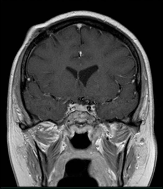
The patient rapidly improved on treatment, with resolution of DI, amenorrhea, and seizures, eventually returning to an almost normal level of health. She completed a year and three months of voriconazole. CSF studies five weeks into treatment showed marked improvement in pleocytosis, and the CSF fungal culture for Blastomyces was negative. Blastomyces urine antigen was negative within six months of treatment. Follow-up MRI of the brain and spine showed decreasing leptomeningeal enhancement without new lesions, consistent with healing (Figure 2). At her follow-up appointment over a month after completing therapy, the patient remained clinically stable, reporting only nausea.
3. Discussion
This case highlights the diagnostic complexity of blastomycosis-associated hypophysitis in a young, otherwise healthy woman without overt pulmonary symptoms or known immunosuppression. The rarity of CNS involvement, let alone pituitary disease, made blastomycosis an unlikely initial consideration. Instead, the clinical picture of a sellar/suprasellar mass with granulomatous inflammation, lymphocytic pleocytosis, and repeatedly negative CSF bacterial, fungal, and AFB cultures, negative urine Blastomyces and Histoplasma antigens, and an initially elevated CSF ACE reasonably led to a working diagnosis of neurosarcoidosis. However, her rapid clinical decline following steroid initiation—including hydrocephalus, cerebral infarcts, and central diabetes insipidus—raised concern for an uncontrolled infectious process. Notably, Blastomyces dermatitidis was ultimately identified on CSF culture more than a month into hospitalization, after immunosuppressive therapy had begun. This timeline suggests that fungal growth may have been unmasked or accelerated following immune modulation. While the patient had been treated with dupilumab in the months preceding illness, her clinical course more closely reflects the vulnerability of even otherwise healthy individuals to invasive CNS blastomycosis, particularly when targeted treatment is delayed.
The diagnosis of CNS blastomycosis is further complicated by the limited sensitivity of available diagnostic tools. CSF culture, while specific, often yields false negatives or delayed results, and growth may take up to five weeks. In a multicenter review of CNS blastomycosis, only 1 of 11 patients had visible yeast on initial CSF examination, and fewer than half had positive CSF cultures, despite confirmed infection [14]. Non-culture testing is likewise imperfect. Urine Blastomyces antigen sensitivity has ranged from 76.3% to 92.9% across cohorts of culture-confirmed cases, with a specificity of 79.3% relative to healthy controls; sensitivity is lower in serum (55.6%) and bronchoalveolar lavage fluid (62.5%) [15]. Cross-reactivity can occur with histoplasmosis, paracoccidioidomycosis, and talaromycosis, limiting specificity. Critically, antigen detection reflects total fungal burden, and sensitivity is lowest in localized or low-burden disease [15]. The performance of antigen testing on CSF remains poorly defined and has not been validated for CNS disease, so a negative result from any compartment cannot be used to exclude CNS blastomycosis. Serum antibody testing directed against the Blastomyces adhesin BAD-1 offers a complementary approach, with a reported sensitivity of 87.8% in proven and probable cases, although cross-reactivity with histoplasmosis persists [15]. Accordingly, no single modality should be relied upon in isolation; current guidance advises combining direct visualization, culture, urine antigen testing, and serum anti-BAD-1 antibody testing, and interpreting them alongside epidemiologic exposure and clinical presentation [14,15].
Despite these diagnostic hurdles, certain overlapping CSF and imaging findings can raise suspicion for fungal CNS infections. CSF pleocytosis is common, particularly in meningeal disease, but is not universal [14]. Leptomeningeal enhancement on MRI is another important radiologic clue that may indicate blastomycosis [14]. Additionally, culture of ventricular fluid may offer greater sensitivity than standard lumbar CSF culture in some cases [14]. When initial diagnostic testing is inconclusive, these findings can support continued investigation and help prevent diagnostic delays.
The recommended treatment for CNS blastomycosis is a lipid formulation of amphotericin B for 4 to 6 weeks, followed by an oral azole for at least 12 months and until resolution of CSF abnormalities [16–18]. Our patient received approximately 4 weeks of liposomal amphotericin B, followed by a year and three months of oral voriconazole. After initiation of antifungal therapy, she gradually improved, with CSF studies showing improvement five weeks into treatment and a follow-up MRI demonstrating decreased leptomeningeal enhancement.
4. Conclusion
Blastomycosis-associated hypophysitis is a rare manifestation of CNS fungal infection that can mimic autoimmune or neoplastic disease. This case demonstrates how such infections may initially present in young, otherwise healthy individuals with minimal systemic symptoms. In endemic regions, clinicians should maintain a high index of suspicion for fungal infections when evaluating atypical pituitary inflammation, particularly in cases that progress and worsen after initiation of steroids or that fail to respond as expected to immunosuppression. Delayed diagnosis remains a challenge due to limited sensitivity of early testing, but persistence in evaluation and timely antifungal therapy can lead to substantial recovery even after a severe course.
Patient perspective: In the patient’s own words: While I was going through this illness, it was the hardest thing I had ever been through. Although I have “recovered”, I still have had to navigate the effects it has left in my everyday life, and I know that I’m going to experience them for the rest of my life. Recovering has given me a different outlook on life, and I’ve tried to remind myself to be patient with finding my new “normal”. I will eternally be grateful for having my mother, family, and team at Rush as my circle of support.
Author Contributions
R.J. drafted the manuscript and performed the literature review. S.S. supervised, revised, and approved the final version. All authors have read and agreed to the published version of the manuscript.
Funding
This research received no external funding.
Institutional Review Board Statement
Not applicable.
Informed Consent Statement
Written informed consent was obtained from the patient to publish this paper and any accompanying images.
Data Availability Statement
No new data were created or analyzed in this study. Data sharing does not apply to this article.
Conflicts of Interest
The authors declare no conflicts of interest.
References
- Miceli, A.; Krishnamurthy, K. Blastomycosis. In StatPearls [Internet] ; StatPearls Publishing: Treasure Island, FL, USA, 2025. Available online: https://www.ncbi.nlm.nih.gov/books/NBK441987/ (accessed on 8 August 2023).
- Smith, D.J.; Williams, S.L.; Endemic Mycoses State Partners Group; Benedict, K.M.; Jackson, B.R.; Toda, M. Surveillance for Coccidioidomycosis, Histoplasmosis, and Blastomycosis—United States, 2019. MMWR Surveill. Summ. 2022, 71, 1–14. [CrossRef]
- Dworkin, M.S.; Duckro, A.N.; Proia, L.; Semel, J.D.; Huhn, G. The epidemiology of blastomycosis in Illinois and factors associated with death. Clin. Infect. Dis. 2005, 41, e107–e111. [CrossRef]
- Centers for Disease Control and Prevention. Case Reporting and National Surveillance for Fungal Diseases. 2023. Available online: https://www.cdc.gov/fungal/php/case-reporting/index.html (accessed on 7 November 2023).
- Pullen, M.F.; Alpern, J.D.; Bahr, N.C. Blastomycosis—Some progress but still much to learn. J. Fungi 2022, 8, 824. [CrossRef]
- Frost, H.M.; Novicki, T.J. Blastomycosis Differential Diagnoses. Medscape Reference. Updated April 2025. Available online: https://emedicine.medscape.com/article/296870-differential (accessed on 8 August 2023).
- Kazmer, A.; El-Baba, R.; Kontosis, A.; Borys, E.; Siddiqui, M. Blastomycosis of the Central Nervous System. J. Radiol. Case Rep. 2024, 17, 13–21. [CrossRef]
- Abrams, E.; Dean, H.J.; Bunge, M.K.; Taback, S.P.; Sellers, E.A. Blastomycosis of the central nervous system in a child: A rare cause of hypopituitarism. J. Pediatr. Endocrinol. Metab. 2011, 24, 775–777. [CrossRef]
- Bush, J.W.; Wuerz, T.; Embil, J.M.; Del Bigio, M.R.; McDonald, P.J.; Krawitz, S. Outcomes of persons with blastomycosis involving the central nervous system. Diagn. Microbiol. Infect. Dis. 2013, 76, 175–181. [CrossRef]
- Morse, H.G.; Nichol, W.P.; Cook, D.M.; Blank, N.K.; Ward, T.T. Central nervous system and genitourinary blastomycosis: Confusion with tuberculosis. West. J. Med. 1983, 139, 99–103. [PubMed] [PubMed Central]
- Mohazab, H.R.; Langer, B.G.; Ramsey, R.G. Intracranial blastomycosis presenting as a suprasellar mass: MR findings. J. Comput. Assist. Tomogr. 1997, 21, 845–847. [CrossRef]
- Mason, A.R.; Cortes, G.Y.; Cook, J.; Maize, J.C.; Thiers, B.H. Cutaneous blastomycosis: A diagnostic challenge. Int. J. Dermatol. 2008, 47, 824–830. [CrossRef]
- Saccente, M.; Woods, G.L. Clinical and laboratory update on blastomycosis. Clin. Microbiol. Rev. 2010, 23, 367–381. [CrossRef]
- Bariola, J.R.; Perry, P.; Pappas, P.G.; Proia, L.; Shealey, W.; Wright, P.W.; Sizemore, J.M.; Robinson, M.; Bradsher, R.W., Jr. Blastomycosis of the central nervous system: A multicenter review of diagnosis and treatment in the modern era. Clin. Infect. Dis. 2010, 50, 797–804. [CrossRef]
- Hage, C.A.; Carmona, E.M.; Epelbaum, O.; Evans, S.E.; Gabe, L.M.; Haydour, Q.; Knox, K.S.; Kolls, J.K.; Murad, M.H.; Wengenack, N.L.; et al. Microbiological Laboratory Testing in the Diagnosis of Fungal Infections in Pulmonary and Critical Care Practice. An Official American Thoracic Society Clinical Practice Guideline. Am. J. Respir. Crit. Care Med. 2019, 200, e45–e67. [CrossRef]
- Castillo, C.G.; Kauffman, C.A.; Miceli, M.H. Blastomycosis. Infect. Dis. Clin. N. Am. 2016, 30, 247–264. [CrossRef]
- Chapman, S.W.; Dismukes, W.E.; Proia, L.A.; Bradsher, R.W.; Pappas, P.G.; Threlkeld, M.G.; Kauffman, C.A. Clinical Practice Guidelines for the Management of Blastomycosis: 2008 Update by the Infectious Diseases Society of America. Clin. Infect. Dis. 2008, 46, 1801–1812. [CrossRef]
- Miller, R.; Assi, M.; AST Infectious Diseases Community of Practice. Endemic fungal infections in solid organ transplant recipients—Guidelines from the American Society of Transplantation Infectious Diseases Community of Practice. Clin. Transplant. 2019, 33, e13553. [CrossRef]