
Abstract
Legionella micdadei is poorly detected by routine urinary antigen and pneumophila-specific polymerase chain reaction assays, contributing to underdiagnosis. We report a case in a 71-year-old woman with Sjögren syndrome who presented with five days of nonproductive cough, fever, and bilateral pulmonary infiltrates unresponsive to outpatient cefpodoxime and azithromycin. Sputum and bronchoalveolar lavage cultures, urine pneumococcal and Legionella antigen tests, L. pneumophila polymerase chain reaction, and Legionella species polymerase chain reaction were all negative; transbronchial cryobiopsy demonstrated organizing pneumonitis without identifiable organisms. The diagnosis was established by plasma microbial cell-free DNA sequencing, which detected L. micdadei.
Highlights
- Legionella micdadei is the second-most commonly identified Legionella species in human disease and disproportionately affects patients with impaired cell-mediated immunity.
- Urinary Legionella antigen testing detects only Legionella pneumophila serogroup 1 and cannot reliably exclude infection caused by non-pneumophila species.
- Legionella polymerase chain reaction (PCR) assays vary in species coverage and may be falsely negative on respiratory specimens, particularly after antibiotic exposure.
- Plasma microbial cell-free DNA (mcfDNA) sequencing can establish a microbiologic diagnosis in culture-negative pneumonia when conventional testing is unrevealing.
- L. micdadei produces nodular infiltrates more often than L. pneumophila and can be weakly acid-fast on modified Kinyoun staining, occasionally leading to misidentification as a mycobacterial species.
- Treatment with a respiratory fluoroquinolone or a macrolide remains first-line therapy; duration extends to 14 to 21 days in immunocompromised hosts.
1. Introduction
Legionella species cause an estimated 2% to 9% of community-acquired pneumonia hospitalizations in the United States, the great majority attributed to Legionella pneumophila serogroup 1 [1]. Non-pneumophila species cause a smaller but clinically important share of disease, and the true incidence is likely underestimated because routine diagnostic testing is biased toward L. pneumophila [2].
Legionella micdadei was first described in 1979 as the Pittsburgh pneumonia agent [3] and is the most frequently isolated non-pneumophila Legionella species [2,4], with a predilection for patients with impaired cell-mediated immunity. Because urinary antigen testing detects only L. pneumophila serogroup 1 and PCR coverage varies by platform [5,6], non-pneumophila legionellosis is frequently underdiagnosed. The following case illustrates unique clinical presentations of, as well as the diagnostic challenge posed by, L. micdadei.
2. Case Presentation
A 71-year-old woman presented with 5 days of nonproductive cough, dyspnea, and subjective fevers and chills. She was previously highly active but had developed marked exertional intolerance over the preceding week. Past medical history was notable for untreated Sjögren syndrome. She denied any exposure to construction and denied any relevant travel history.
A chest radiograph obtained as an outpatient two days before admission was clear. One day before admission, repeat imaging demonstrated bilateral pulmonary infiltrates, and she was started on cefpodoxime and azithromycin as an outpatient, without clinical improvement.
On admission, temperature was 30.4 °C, heart rate 100 beats per minute, respiratory rate 22, blood pressure 136/73 mmHg, and oxygen saturation 96% on room air. She appeared well. Cardiac and pulmonary examinations were unremarkable, and there were no skin findings.
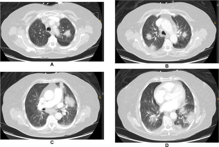
Laboratory studies were notable for sodium 130 mmol/L, glucose 155 mg/dL, aspartate aminotransferase 78 U/L, alanine aminotransferase 86 U/L, total bilirubin 1.7 mg/dL, albumin 3.3 g/dL, and platelets 107 × 109/L. The remainder of the chemistry panel and complete blood count were within normal limits. Autoimmune serologies were consistent with her known Sjögren syndrome (antinuclear antibody 1:160 speckled, SSA IgG 17). Contrast-enhanced computed tomography (CT) of the chest demonstrated multifocal airspace disease, mediastinal and hilar lymphadenopathy, and no evidence of pulmonary embolism (Figure 1).
Blood cultures, HIV serology, urine pneumococcal antigen, urine Legionella antigen, Quantiferon-TB, and transthoracic echocardiogram were all negative. A comprehensive fungal serologic panel including Aspergillus galactomannan, Blastomyces antibody and antigen, Coccidioides antibodies, Cryptococcus antigen, and Histoplasma antibody and urine antigen was negative. Bronchoscopy with bronchoalveolar lavage was performed; bacterial, fungal, and acid-fast bacilli cultures were negative, as were L. pneumophila PCR and qualitative Legionella species PCR on the bronchoalveolar lavage (BAL) specimen. Transbronchial cryobiopsy of left and right upper lobe nodules demonstrated organizing pneumonitis with chronic inflammation and fibrosis, negative for malignancy, with negative Gomori methenamine silver (GMS) staining for fungi.
The patient remained clinically stable and was never hypoxic. She completed 3 days of azithromycin and was transitioned from ceftriaxone to amoxicillin-clavulanate to complete a 7-day course of empiric therapy. Plasma microbial cell-free DNA sequencing (Karius test) subsequently returned with detection of Legionella micdadei (Tatlockia micdadei) at 14,898 molecules per 100 nanoliters, flagged as an obligate pathogen alert result. She was further prescribed 7 days of levofloxacin. She remained well at her 2-week follow-up without recurrence of symptoms. Repeat chest CT at week 4 showed complete resolution.
3. Discussion
3.1. Legionella micdadei and Non-Pneumophila Legionellosis
L. micdadei is a fastidious, gram-negative bacillus that replicates intracellularly within free-living amoebae in aqueous environments and within alveolar macrophages once aerosolized and inhaled by a susceptible host [7]. Unique among Legionella species, it is weakly acid-fast on modified Kinyoun staining and may be misidentified as a mycobacterium in tissue [2,4]. The organism grows on buffered charcoal yeast extract (BCYE) agar over 3 to 5 days at 35 °C. L. longbeachae, L. bozemanii, and L. dumoffii are other clinically encountered non-pneumophila species, with geographic variation in distribution [2].
Risk factors for L. micdadei pneumonia overlap with those for other Legionella species and include age greater than 50 years, smoking, chronic heart or lung disease, diabetes, and end-stage renal disease [2,7]. Patients with impaired cell-mediated immunity, including solid organ transplant recipients, those on chronic corticosteroids, and those with hematologic malignancy, are at particularly elevated risk [2,4]. Sjögren syndrome is associated with subtle deficits in cellular immunity and B-cell hyperactivity and may have contributed to host susceptibility in this case [8].
3.2. Limitations of Conventional Diagnostic Testing
This case illustrates two recurring challenges in the diagnosis of legionellosis. First, urinary antigen testing, the most widely available and rapid Legionella assay, is designed to detect the lipopolysaccharide antigen of L. pneumophila serogroup 1. Reported sensitivity is approximately 90% for L. pneumophila serogroup 1, 60% for other L. pneumophila serogroups, and as low as 5% for non-pneumophila species including L. micdadei [6]. A negative urinary antigen therefore cannot exclude legionellosis when clinical suspicion is high.
Second, Legionella nucleic acid amplification tests vary considerably in target coverage. Some commercial assays detect only L. pneumophila, while broader Legionella species PCRs use 16S rRNA or mip gene targets and should, in principle, identify L. micdadei [5]. In this patient, both pneumophila-specific and species-level PCR on BAL fluid were negative, likely reflecting low organismal burden, prior antibiotic exposure, and assay-specific limitations [5,6]. Culture yield similarly falls once antibiotics have been administered, further limiting confirmatory diagnosis after empiric therapy [2].
3.3. Microbial Cell-Free DNA Sequencing in Culture-Negative Pneumonia
Plasma microbial cell-free DNA (mcfDNA) sequencing detects fragments of pathogen-derived DNA circulating in plasma and can identify more than 1000 bacteria, viruses, fungi, and parasites from a single specimen [9]. In its original validation study, mcfDNA sequencing demonstrated approximately 93% agreement with composite reference methods for pneumonia pathogen identification, and subsequent clinical impact studies have shown the greatest yield in immunocompromised hosts, culture-negative pneumonia, and infections caused by atypical or fastidious organisms [9,10]. In this patient, the L. micdadei signal was high (14,898 molecules per 100 nanoliters), and the assay classified Legionella as an obligate pathogen unlikely to represent commensal or contaminant DNA at any concentration. Combined with the compatible clinical and radiographic picture and the absence of an alternative pathogen, the result was interpreted as causal.
3.4. Radiographic and Clinical Features of L. micdadei Pneumonia
In contrast to L. pneumophila, which classically produces lobar or patchy alveolar infiltrates, L. micdadei more commonly presents with nodular opacities and, in immunocompromised hosts, with cavitating nodules that may be mistaken for malignancy, septic emboli, fungal pneumonia, or tuberculosis (Figure 1) [2,4]. In this patient, the initial radiographic differential included these entities, prompting transbronchial cryobiopsy, which demonstrated organizing pneumonitis, a nonspecific reaction pattern consistent with but not diagnostic of Legionella pneumonia [2].
Extrapulmonary laboratory abnormalities frequently accompany legionellosis and were present here, including hyponatremia, mild transaminitis, hypoalbuminemia, and thrombocytopenia. These findings are nonspecific but support the diagnosis in the right clinical context [2,7].
3.5. Treatment Considerations
Antimicrobial treatment of L. micdadei pneumonia mirrors that of L. pneumophila. Respiratory fluoroquinolones (levofloxacin or moxifloxacin) and macrolides (azithromycin) are first-line, with comparable efficacy in observational and randomized studies [11]. Beta-lactams have no clinically useful activity against Legionella because of intracellular replication and beta-lactamase production [2, 11].
Duration of therapy is typically 7 to 10 days in immunocompetent hosts and 14 to 21 days in immunocompromised patients or those with severe or complicated disease [11].
3.6. Implications for the Diagnostic Algorithm
Approximately half of community-acquired pneumonia cases remain microbiologically undefined despite contemporary testing [12]. In hospitalized patients who fail empiric outpatient therapy, multimodal testing combining culture, urinary antigen, respiratory PCR, and bronchoscopy with BAL offers the greatest yield [12]. When this evaluation is unrevealing and clinical suspicion for an atypical or fastidious pathogen remains, plasma mcfDNA sequencing is a reasonable next step, balanced against cost, turnaround time, and the risk of detecting clinically irrelevant organisms [9,10].
3.7. Conclusion
L. micdadei is a clinically important cause of community-acquired pneumonia that is poorly detected by urinary antigen testing and may be missed by Legionella-targeted PCR, particularly after antibiotic exposure. In patients with pneumonia unresponsive to standard empiric therapy, especially those with nodular infiltrates or impaired cell-mediated immunity, a high index of suspicion for non-pneumophila Legionella should be maintained, and plasma microbial cell-free DNA sequencing should be considered when conventional testing is unrevealing.
Funding
This research received no external funding.
Acknowledgments
The author used Claude for content assistance, specifically to create an initial draft from prior personal presented material. The manuscript was reviewed and the author revised the material generated and takes full responsibility for the content of this publication.
Conflicts of Interest
The author declares no conflict of interest.
References
- Cunha, B.A.; Burillo, A.; Bouza, E. Legionnaires’ disease. Lancet 2016, 387, 376–385. [CrossRef] [PubMed]
- Muder, R.R.; Yu, V.L. Infection due to Legionella species other than L. pneumophila. Clin. Infect. Dis. 2002, 35, 990–998. [CrossRef] [PubMed]
- Pasculle, A.W.; Feeley, J.C.; Gibson, R.J.; Cordes, L.G.; Myerowitz, R.L.; Patton, C.M.; Gorman, G.W.; Carmack, C.L.; Ezzell, J.W.; Dowling, J.N. Pittsburgh pneumonia agent: Direct isolation from human lung tissue. J. Infect. Dis. 1980, 141, 727–732. [CrossRef] [PubMed]
- Fang, G.D.; Yu, V.L.; Vickers, R.M. Disease due to the Legionellaceae (other than Legionella pneumophila). Historical, microbiological, clinical, and epidemiological review. Medicine 1989, 68, 116–132. [CrossRef] [PubMed]
- Mercante, J.W.; Winchell, J.M. Current and emerging Legionella diagnostics for laboratory and outbreak investigations. Clin. Microbiol. Rev. 2015, 28, 95–133. [CrossRef] [PubMed]
- Helbig, J.H.; Uldum, S.A.; Bernander, S.; LuüK, P.C.; Wewalka, G.; Abraham, B.; Gaia, V.; Harrison, T.G. Clinical utility of urinary antigen detection for diagnosis of community-acquired, travel-associated, and nosocomial legionnaires’ disease. J. Clin. Microbiol. 2003, 41, 838–840. [CrossRef] [PubMed]
- Phin, N.; Parry-Ford, F.; Harrison, T.; Stagg, H.R.; Zhang, N.; Kumar, K.; Lortholary, O.; Zumla, A.; Abubakar, I. Epidemiology and clinical management of Legionnaires’ disease. Lancet Infect. Dis. 2014, 14, 1011–1021. [CrossRef] [PubMed]
- Mariette, X.; Criswell, L.A. Primary Sjögren’s Syndrome. N. Engl. J. Med. 2018, 378, 931–939. [CrossRef] [PubMed]
- Blauwkamp, T.A.; Thair, S.; Rosen, M.J.; Blair, L.; Lindner, M.S.; Vilfan, I.D.; Kawli, T.; Christians, F.C.; Venkatasubrahmanyam, S.; Wall, G.D.; et al. Analytical and clinical validation of a microbial cell-free DNA sequencing test for infectious disease. Nat. Microbiol. 2019, 4, 663–674. [CrossRef] [PubMed]
- Hogan, C.A.; Yang, S.; Garner, O.B.; Green, D.A.; Gomez, C.A.; Bard, J.D.; Pinsky, B.A.; Banaei, N. Clinical Impact of Metagenomic Next-Generation Sequencing of Plasma Cell-Free DNA for the Diagnosis of Infectious Diseases: A Multicenter Retrospective Cohort Study. Clin. Infect. Dis. 2021, 72, 239–245. [CrossRef] [PubMed]
- Garin, N.; Genné, D.; Carballo, S.; Chuard, C.; Eich, G.; Hugli, O.; Lamy, O.; Nendaz, M.; Petignat, P.-A.; Perneger, T.; et al. β-Lactam monotherapy vs β-lactam-macrolide combination treatment in moderately severe community-acquired pneumonia: A randomized noninferiority trial. JAMA Intern. Med. 2014, 174, 1894–1901. [CrossRef] [PubMed]
- Metlay, J.P.; Waterer, G.W.; Long, A.C.; Anzueto, A.; Brozek, J.; Crothers, K.; Cooley, L.A.; Dean, N.C.; Fine, M.J.; Flanders, S.A.; et al. Diagnosis and Treatment of Adults with Community-acquired Pneumonia. An Official Clinical Practice Guideline of the American Thoracic Society and Infectious Diseases Society of America. Am. J. Respir. Crit. Care Med. 2019, 200, e45–e67. [CrossRef] [PubMed]